


Varicose veins of the lower extremities are characterized by the expansion of the superficial veins of the legs, which accompanies the violation of blood flow in them and the failure of the valves. As a result, the veins increase in length and diameter, acquire a serpentine, cylindrical or saccular appearance, although there is also a mixed manifestation of the listed deformities.
Characteristics of the venous system.
The appearance and development of varicose veins is directly related to the venous system of the legs, which consists of:
- saphenous veins: small and large;
- deeply located veins (in the lower leg and thigh);
- perforating veins, which are the connecting link of the two previous systems.
Normally, 90% of the blood is transported to the lower extremities through deep veins and the remaining 10% through superficial veins. When it returns to the side of the heart, this mechanism is supported by valves in the walls of the veins. When the next portion of blood arrives, they strike to prevent it from moving up and down under the influence of gravitational force. Muscle contractions push blood further toward the heart, allowing normal blood flow.
With a prolonged stay of a person in an upright position, stagnation of the blood can develop, which increases the pressure in the veins and causes an increase in their diameter. This process causes incomplete closure of the valve leaflets, as a result of which the blood flow is disturbed with its reverse flow from the heart - reflux.
Valves in deep veins are more likely to be affected as they carry the greatest amount of blood and therefore experience the maximum load. To lower the high pressure in them, part of the blood is transported by perforated veins to the superficial ones, which were not originally intended for a large volume. Such a load on the walls of the veins leads to their expansion and the formation of varicose veins.
At the same time, the blood enters the deep veins without stopping, but due to the violation of their functions and the normal activity of the leaflets of the perforated veins, the blood is redistributed to the superficial vessels. As a result, chronic varicose veins develop, which over time are accompanied by painful sensations, edema, and trophic ulcers.
Causes of the disease
Previously, one of the main causes of varicose veins was called a hereditary factor, but today this theory has been disproven. Of course, it is possible to trace the frequent manifestations of the disease in some families, but this is more likely due to the peculiarities of life that are taught in the family: food culture, passive rest, sedentary work, etc.
The development of varicose veins is based on the presence of reflux in the venous system, when blood circulates through the veins in the opposite direction. Additional blood transport from deep veins to superficial veins is possible due to congenital or acquired degenerative disease of the valve apparatus. This causes the superficial vessels to overfill with blood and their distention when the venous ganglia form.
One of the fundamental reasons for the development of varicose veins is considered an unhealthy diet, which in some cases leads to obesity. These people move little, eat mainly highly processed foods, and the proportion of plant fiber in the diet is minimized. After all, it is they who are involved in strengthening the walls of the veins and blood vessels and prevent long-term chronic constipation, which greatly increases intra-abdominal pressure and thus provokes varicose veins. It is observed that an increase in body weight of more than 20% increases the risk of disease five times.
The main provoking factor for women is having a child, while the risks of varicose veins increase with each subsequent pregnancy. Severe weight gain and an enlarged uterus put a lot of pressure on the legs, which are stagnant. This situation is aggravated by the constantly growing intra-abdominal pressure and the action of the hormone progesterone, which affects the state of the elastic fibers in the walls of the blood vessels.
Other factors that cause varicose veins of the lower extremities include:
- a sedentary lifestyle, being on your feet during the day (for example, hairdressers), long flights or long trips. All this leads to stagnant processes in the lower extremities, when blood accumulates in the superficial veins and is poorly transported to the heart;
- sometimes increases the risk of developing varicose veins for women, the use of uncomfortable and tight shoes, especially models with high heels;
- corsets and tight underwear tighten the inguinal veins and increase intra-abdominal pressure, which is a direct prerequisite for varicose veins;
- High blood pressure;
- smoking, which indirectly leads to a thinning of the blood vessel walls.
Disease classification
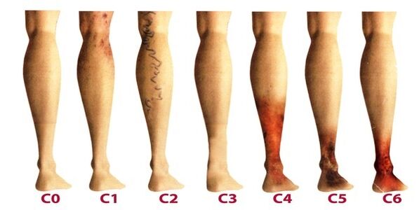
Varicose veins of the lower extremities are classified according to the prevalence of venous lesions, their location, and the presence of pathological reflux, which is characterized by impaired blood flow. There are 4 forms of varicose veins:
- intracutaneous and subcutaneous (segmental) varicose veins, in which there is no pathological outflow of venous blood;
- Segmental varicose veins, when reflux occurs through perforating or superficial veins;
- a common form of varicose veins, in which reflux occurs through the perforating and superficial veins at the same time;
- Varicose veins are characterized by deep vein reflux.
After the varicose veins of the lower extremities become chronic, phlebology considers its three degrees:
- Transient edema, which occurs periodically against the background of "heavy legs" syndrome.
- Persistent and persistent edema. Hyperpigmentation and eczema may appear.
- Venous ulcer of a trophic nature.
The last degree is the most difficult to treat, as it requires the preliminary removal of inflammation and healing of the skin tissues.
Stages and symptoms
The disease develops very slowly, sometimes more than a dozen years pass, until the symptoms that appear will force the patient to seek the advice of a phlebologist. In the initial stages of varicose veins, its manifestations are often attributed to fatigue, age, or other reasons. In order to fully consider the symptoms of the disease, its manifestations are classified according to the stages of varicose veins:
- The first stage begins to manifest itself most often at an early age: after the age of 20, when there is a feeling of heaviness in the legs, edema may appear, which completely disappears overnight. On the inside of the lower leg, you can see an enlarged vein, which is manifested by a bulging bump of the skin. At this stage, many people notice tiny spider veins. In general, the symptoms are subtle and rarely receive the attention it deserves.
- The second stage is characterized by an increase in the external manifestation of the dilated vein. The disease is already developing against the background of the pathological work of the venous valves, therefore, the saphenous veins markedly increase in size, and their elongation can also be noted. Most often there is heaviness and burning in the legs, they quickly tire from long walks.
- The disease is already becoming chronic due to the constant imbalance in venous blood outflow. At night, patients suffer from edema near the ankle, which can be very intense. There is heaviness in the legs and cramps may occur at night.
- In the absence of treatment in the previous stages, chronic insufficiency of the functioning of the venous system adversely affects metabolic processes in the skin, the areas in the lower leg are especially affected. Darkening of the skin is visible near the ankle: hyperpigmentation, thickens and swells over time. The condition described is called lipodermatosclerosis. If at this time you do not start therapy regarding the venous system, soon trophic ulcers will begin to form.
- The fifth stage is accompanied by numerous trophic ulcers, some of them periodically heal with the formation of scars.
- In the area of long-lasting trophic changes, extensive ulcers open. This condition requires urgent active therapy, aimed at both the treatment of varicose veins and the healing of skin ulcers.
Diagnostics
An external examination of the lower extremities in vertical and horizontal positions of the body, palpation of the veins and a preliminary assessment of the stage of the disease is performed. The patient is sent for a general blood test, which allows him to study the picture of the disease more closely:
- at the platelet level, a predisposition to thrombosis will be reflected;
- the level of hemoglobin, as well as the number of red blood cells, indicate the degree of blood clotting;
- By increasing the level of leukocytes, it can be judged on the inflammation, which helps to diagnose thrombophlebitis more quickly.
Be sure to examine the venous system of the legs, for which there are many methods:
- Ultrasound Dopplerography - USDG;
- phlebography;
- CT phlebography;
- duplex angioscan - USAS;
- phleboscintiography;
- photoplesmography;
- phlebomanometry and the like.
In practice, patients are more often prescribed USAS and USG, as they help to fully study the venous system of the legs and identify degenerative areas. The rest of the methods can be additionally prescribed if the ultrasound examination did not provide a complete picture of the disease. Some of these methods can have complications such as venous thrombosis, perforation of the vessel wall with a catheter, and allergy to the contrast medium. Consider the most commonly practiced techniques in phlebology:
- USAS allows evaluating the anatomical, hemodynamic and functional pathologies of the venous bed. The data obtained is subject to computer processing, after which the venous system model can be viewed on video or printed on paper.
- High-precision Doppler ultrasound determines the patency of superficial and deep veins, the speed of blood flow. Doppler ultrasound allows evaluating the functioning of the valve apparatus.
After an extensive diagnosis, the doctor draws up a patient's flebocard, which allows him to determine the damaged segments of the venous system, their degree and length. After that, a suitable treatment is selected.
Treatment
It is performed comprehensively and is determined based on the symptoms, the degree of development of the disease and the results of the study. In the initial stages, conservative therapy is prescribed, which consists of:
- Drug treatment when a group of drugs is prescribed:
- antioprotectors and phlebotonics;
- anticoagulants;
- disaggregating
- topical preparations (ointments, gels);
- anti-inflammatory drugs.
- Elastic compression, for which compression stockings or bandages are used (rarely). It allows you to dose the compression of the muscles, prevents stagnant processes, improves blood flow through the vessels. When wearing such an underwear, there is the effect of artificially maintaining vascular tone.
- Physiotherapeutic methods, among which the best treatment results were shown by electrophoresis, diadynamic currents, laser radiation and magnetic field.
- Feasible physical activity, which should be done only in compression underwear (except for swimming). Biking, swimming, jogging is recommended. The phlebologist selects an individual set of exercises for the lower extremities, which will train the vessels of the legs every day.
In addition, patients are advised to perform contrasting five-minute procedures in the shower every night, alternately changing from warm to cold water. Such manipulations improve blood flow and tone blood vessels.
It is important at the beginning of treatment to identify the factor that causes the disease to be able to influence it effectively. And patients who are at risk should visit a phlebologist every 2 years for a preventive examination and an ultrasound examination of the veins in the legs.
When conservative treatment is unsuccessful or varicose veins are seen at an advanced stage, surgical intervention is used. Today, varicose veins can be completely cured thanks to the following methods:
- Phlebectomy. The essence of the operation is to remove the main trunks of the superficial vein to eliminate the pathological discharge of blood. Perforating veins are often ligated for the same purpose.
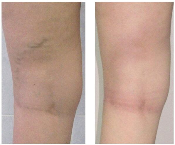
- Sclerotherapy. It consists of the introduction of a sclerosant in the affected area of the vein, which leads to the connection of its walls. Recently, they began to actively use foam sclerosing agent for the same purposes according to the technology -. Blood flow through the defective area is stopped and the cosmetic defect in the form of protruding nodules is removed. After such an intervention, no scars remain, all manipulations are carried out on an outpatient basis without a subsequent hospital stay. But sclerotherapy is used only for the fusion of small branches of the venous trunks.
- Laser coagulation. With the help of a laser beam, the marked section of the vein, the walls of which come together, is heated and the flow of blood through it is stopped. But this technique is indicated only for veins with an expansion diameter of less than one centimeter.
Prevention
Preventive measures can be both primary, aimed at preventing the development of varicose veins, and secondary, when required to reduce the risk of relapse after surgery or to prevent the worsening of the course of the disease. Helpful tips:
- lead an active lifestyle without a heavy load on the legs: swimming, walking, cycling;
- watch your weight;
- keep both legs raised more often;
- do not wear tight underwear and heels larger than 4 centimeters;
- wear orthotic inserts;
- take a contrast shower;
- do five-minute preventative leg exercises every day;
- wear compression stockings for long walks.
If you notice the slightest suspicion of varicose veins - prominent nodules on the legs, swelling, heaviness, do not postpone the visit to the phlebologist. In fact, over time, this insidious disease can lead to many complications, such as thrombophlebitis and thrombosis.